Publikace v časopise Inorganic Chemistry Communications
Structural variations in copper(II) amine-bisphenolate complexes: Evaluation of in vitro antiproliferative activity against human cancer and normal cells
Authors: Anssi Peuronen, Pia Damlin, Ján Vančo, Zdeněk Dvořák, Zdeněk Trávníček, Ari Lehtonen
Full-text: https://doi.org/10.1016/j.inoche.2025.115024
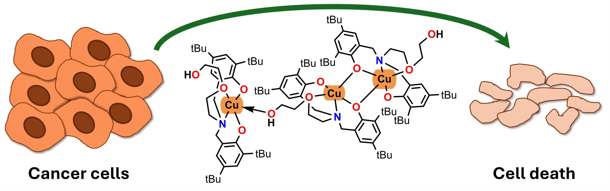
Three copper(II) complexes with variously substituted amine-bisphenolates (H2L1, H2L2 and H2L3) have been prepared. Variation in the composition and structure of the free ligands resulted in the formation of three structurally distinct copper(II) complexes: dinuclear 1, mononuclear 2 and trinuclear 3. Various physical techniques were used to characterise the complexes, including single-crystal X-ray analysis and variable-temperature magnetic susceptibility measurements (5–297 K). The compounds were evaluated for their in vitro anti-proliferative effects against three human cancer cell lines (ovarian A2780 and A2780R, breast MCF7) and normal HaCaT cells. The results showed that both the free ligands and the complexes exhibit strong-to-moderate cytotoxicity. 2 and 3 are significantly more effective against A2780, A2780R and MCF7 cells than the metallodrug cisplatin. The cytotoxicity of complexes 1–3 is bound to the cytotoxicity of the free ligands and remains almost unchanged over 24, 48 and 72 h. The copper accumulation in A2780 cells was studied by ICP-MS over 2–72 h of co-incubation of 1–3. Complex 1 caused the highest uptake of copper into A2780 cells, reaching up to 100 times higher Cu concentration compared to untreated cells, while 2 and 3 showed only ca 5–10-fold increase of Cu uptake in A2780 cells. No apparent signs of hydrolysis of 1–3 in a MeOH/water mixture were observed in mass spectrometry experiments even after 72 h of standing at laboratory temperature. The mass spectrometry-based interaction studies of 1–3 with L-cysteine (Cys) and reduced glutathione (GSH) did not show direct evidence of the interaction product formation. Only the signals, corresponding to the free ligands were identified in mass spectra after 24 h and 72 h of incubation.